Updated January 12, 2023
Magnetic Resonance Imaging(MRI)
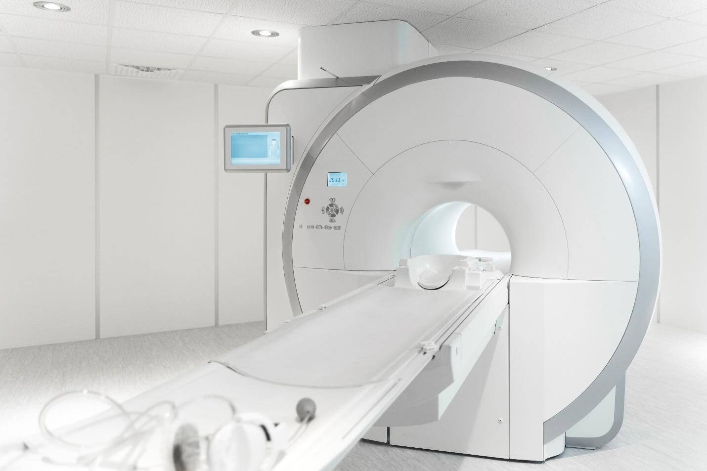
MRI (Magnetic Resonance Imaging) is a kind of visualizing device that produces detailed images of the body’s organs and tissues without radiation. It diagnoses various medical conditions, including cancer, heart, and vascular diseases, central nervous system diseases, and musculoskeletal disorders. An MRI employs an intense magnetic field and radio waves to create comprehensive images of the inside of the body. The images can find abnormalities in the body and track how diseases get worse.
History
Magnetic Resonance Imaging is a diagnostic medical device that produces detailed images of the body’s internal organs and structures using powerful magnetic fields and radio waves. In the 1970s, a team at the University of Aberdeen in Scotland was the first to work on this technology. The inventors, led by Sir Peter Mansfield, used the principles of nuclear magnetic resonance (NMR) to create the images. In 1980, Atkinson Morley Hospital in London installed the first MRI machine.
Since then, it has become one of the most widely used medical imaging techniques. It has revolutionized the diagnosis and treatment of many diseases, including cancer, stroke, heart disease, and neurological disorders. Its non-invasive nature makes it an ideal tool for detecting and monitoring conditions without exposing the patient to radiation or contrast agents. scanners have become increasingly sophisticated, incorporating new technologies such as contrast agents and specialized coils. Today, it is a reliable and invaluable tool for medical diagnosis.
Mechanism
MRI is a non-invasive screening procedure that creates intricate scans of the organs and structures within the body using radio waves, a strong magnetic field, and a computer. The magnetic field aligns the magnetic moments of the atoms in the body, while the radio waves cause the aligned spins to produce a rotating magnetic field. The receiver portion of the MRI detects this rotating magnetic field, which then creates an image of the scanned area.
Diagnostics
MRI is a tool used by medical professionals to diagnose various conditions and diseases. It scans detect tumors, blood vessel disease, stroke, brain, and spinal cord injuries, multiple sclerosis, Alzheimer’s disease, and damaged cartilage. Scans can also provide detailed images of the body’s organs, soft tissues, and bones to aid in diagnosing a wide range of medical conditions. Scans are a non-invasive, safe way to diagnose many medical conditions.
Usage by Organ
It is routinely used to examine the brain, heart, lungs, kidneys, liver, pancreas, and spine. It can detect tumors, diagnose and monitor diseases, evaluate the effects of medical treatments, and assess the health of organs. It is beneficial for imaging soft tissues and organs that don’t appear well on X-rays, such as the brain, spinal cord, and ligaments.
Specialized Configurations
1. High-Field MRI: They are typically used in clinical practice because they provide higher-resolution images and a better signal-to-noise ratio.
2. Open MRI Systems: Have a wider bore, allowing for more comfort and less claustrophobia for patients.
3. Functional MRI (fMRI): fMRI measures brain activity by monitoring variations in blood circulation connected with neural activation.
4. Diffusion Tensor Imaging (DTI): DTI is a non-invasive MRI technique used to measure the diffusion of water molecules in biological tissues.
5. Magnetic Resonance Angiography (MRA): MRA is a type of MRI that uses magnetic fields to produce detailed images of vessels and organs.
6. Magnetic Resonance Spectroscopy (MRS): MRS is a technique used to measure the concentrations of various molecules in a sample.
7. Multi-Parametric MRI: Multi-parametric MRI combines multiple techniques to provide better diagnostic information in a single scan.
8. Ultra-High Field MRI: Ultra-high field MRI systems are the most powerful, and scholars use these for research purposes.
Safety Measures to be Taken while Using MRI
1. Wear a Lead Apron: It is essential to wear a lead apron while undergoing an MRI to protect the body from the solid magnetic radiation emitted.
2. Limit Movement: When undergoing, it is essential to limit movement as much as possible. Any sudden movement may cause the person to become injured.
3. Follow Safety Instructions: It is essential to follow all safety instructions provided by the technician or doctor before the MRI. It includes removing all metal objects from the body and remaining still during the procedure.
4. Monitor Breathing: It is essential to monitor breathing during the MRI as the strong magnetic fields can cause a person to experience difficulty breathing or even pass out.
5. Keep Jewelry Away: It is essential to remove all jewelry and metal objects before entering the MRI room. A strong magnetic field can pick up these objects and cause injury.
Final Thoughts – MRI
MRI is a powerful tool for diagnosing and monitoring various medical conditions. It is a non-invasive and safe imaging modality used in medicine for decades. It has been used to analyze different situations, from cancer and stroke to musculoskeletal injuries. It has also been used in research to study the structure and function of the human body. As technology advances, MRI technology improves and provides more detailed images with less radiation exposure. Although it can be expensive, its accuracy and detail make it a valuable tool in medical diagnosis.