


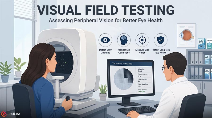
Most people think of eyesight as the ability to read letters, recognize faces, or see objects directly in front of them. Yet central vision is only one part of the visual system. Peripheral, or side, vision helps people notice movement, navigate crowded spaces, drive, avoid obstacles, and maintain awareness of what is happening beyond the point they are directly looking at. Changes in peripheral vision may develop slowly. Because the brain can adapt and the other eye may compensate, a person may not immediately notice that part of the visual field has become less sensitive. This is one reason visual field testing is an important component of eye care, particularly when clinicians are evaluating or monitoring conditions that can affect the optic nerve or visual pathways.
Key Point:
Peripheral vision can change gradually and may not be obvious in daily life. Visual field testing helps measure side vision, but online screening tools do not replace a comprehensive examination by a qualified eye-care professional.
What is the Visual Field?
The visual field is the entire area a person can perceive while looking straight ahead and keeping the eyes still. It includes central vision, which provides detail, and peripheral vision, which covers the wider area surrounding the center of sight. A visual field defect may appear as a dim area, an isolated blind spot, reduced sensitivity, or a broader loss of side vision.
Visual field changes do not all have the same cause or pattern. Eye conditions, optic nerve damage, retinal disease, neurological disorders, eyelid position, fatigue, poor test concentration, and even an incorrect test setup can influence results. For that reason, clinicians interpret a field test alongside an eye examination, medical history, eye-pressure measurements, optic nerve imaging, and previous test results.
Why is Peripheral Vision Easy to Overlook?
Central vision receives attention because it is used for reading, screens, and fine detail. Peripheral vision is less precise but essential for orientation and safety. A person with a developing field defect may still read a standard eye chart well, especially in the early stages of certain conditions.
The National Eye Institute explains that glaucoma can cause loss of side vision and that a comprehensive glaucoma examination may include a visual field test to check peripheral vision. The American Academy of Ophthalmology likewise notes that visual field testing can identify areas of vision loss outside the central field. These findings do not mean that every visual field abnormality is due to glaucoma, but they show why side vision needs to be assessed separately from visual acuity.
How Does Visual Field Testing Work?
Visual field tests measure whether a person can detect visual targets in different locations while maintaining focus on a central point. The exact method varies, but the goal is generally to map sensitivity across the field of vision.
1. Confrontation Testing
A clinician covers one of the patient’s eyes and compares the patient’s ability to notice fingers, movement, or targets in different areas. This is quick but less detailed than automated testing.
2. Automated Static Perimetry
A machine presents small lights at different positions and brightness levels. The patient presses a button when they see a light. Software then creates a map showing sensitivity and possible defects.
3. Kinetic Perimetry
A target moves from an area where it cannot be seen toward an area where it becomes visible. This can help outline the boundaries of the visual field.
4. Screening Tests
Simplified clinic-based or digital tests may identify possible abnormalities that warrant further assessment. Healthcare professionals design them for triage or awareness rather than a final diagnosis.
What Can Affect Test Accuracy?
Visual field testing requires concentration. The patient must maintain fixation on the fixation point, respond consistently, and avoid searching for each stimulus. Results may be less reliable when the person is tired, anxious, distracted, unfamiliar with the procedure, or physically uncomfortable. Other influences include an incorrect prescription for the testing distance, a drooping eyelid, a small pupil, dry eyes, lens rim artifacts, delayed responses, or pressing the response button when no target was presented.
Automated systems usually report reliability indicators, but these indicators must still be interpreted in context. A single unusual result therefore does not always establish a lasting defect. Eye-care professionals often compare repeated tests to determine whether an abnormality is reproducible and whether it is stable, improving, or progressing over time.
The Role of Online Visual Field Screening
Digital tools can increase awareness of peripheral vision and may provide an accessible first check. For example, an online visual field test can help users explore whether they notice targets across different parts of the screen. This may be useful for education, self-observation, or deciding whether to arrange a professional examination.
However, screen size, viewing distance, display brightness, room lighting, device calibration, browser scaling, user technique, and the absence of clinical supervision can all affect the accuracy of online results. A home test cannot examine the optic nerve, measure eye pressure, assess the retina, or rule out neurological causes. You should therefore present it as a screening or awareness tool, not as a substitute for medical diagnosis.
When is Professional Assessment Important?
Routine eye examinations are important even when vision seems normal. The National Eye Institute explains that a dilated eye exam allows a specialist to look for indications of illness in the retina and optic nerve. An appropriate examination schedule can be recommended based on age, family history, eye pressure, optic nerve appearance, existing eye disease, medications, systemic health, and previous findings.
Prompt assessment is especially important after a sudden vision change. New blind areas, a curtain-like shadow, sudden flashes with an increase in floaters, severe eye pain, marked redness, headache with visual symptoms, weakness, facial drooping, speech difficulty, or sudden loss of part of the visual field may require urgent medical attention. People should not rely on an online screening tool when symptoms are sudden, severe, or rapidly changing.
How to Prepare for a Visual Field Test?
Before your visual field testing appointment, follow these recommendations:
- Bring the glasses normally used for near or distance vision, as instructed by the clinic.
- Tell the examiner about fatigue, discomfort, dry eyes, drooping eyelids, or difficulty maintaining fixation.
- Keep looking at the central target rather than searching around for lights.
- Only press the response button when you detect a stimulus; excessive guessing can reduce reliability.
- Ask for a brief pause if concentration declines or the eye becomes uncomfortable.
- Remember that a learning effect is common, so later tests may be more consistent than the first one.
Understanding Results Over Time
Visual field reports contain maps, numerical sensitivity values, summary indices, and statistical comparisons. These reports can appear definitive, but their interpretation is not based on a single number. Clinicians review the shape and location of any defect, test reliability, correspondence with optic nerve or retinal findings, and changes across repeated examinations.
Monitoring over time is particularly important because a stable defect and a progressing defect may require different management. Consistent testing conditions and repeated measurements help clinicians distinguish true change from normal variability.
Final Thoughts
Peripheral vision is an essential but sometimes overlooked part of sight. Visual field testing provides information that a standard letter chart cannot, helping clinicians evaluate how well different areas of vision are functioning. Digital screening can improve awareness and access, provided users clearly understand its limitations. The most responsible approach is to treat online testing as an educational first step. Results that are abnormal, inconsistent, or accompanied by symptoms should be discussed with an optometrist or ophthalmologist. Regular professional eye examinations remain the appropriate way to diagnose eye disease and protect vision over time.
Medical Disclaimer
This content is intended for educational purposes only and should not be considered medical advice or treatment. Anyone concerned about vision changes should consult a qualified eye-care professional.
Recommended Articles
We hope this guide on visual field testing has improved your understanding of peripheral vision and eye health. Explore these recommended articles for more insights and health tips.